The Spine

An animal’s spine is a very complex structure consisting of bones, ligaments, muscles and nerves. It fulfills various functions in the body providing:
- A framework of support
- Attachment for many muscles
- Protection of the spinal cord
- Protection of internal organs
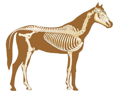
An animal’s spine consists of bones called vertebrae. A horse’s spine is made up of 7 cervical vertebrae, 18 thoracic vertebrae (that connect with the ribs), 6 lumbal vertebrae, 5 sacral vertebrae (which are fused, creating the sacrum) and 16 to 18 coccygeal vertebrae. Where one vertebra connects with another it is called a joint (there are approximately 200 joints in a horse’s spine). Joints are held together by a vast number of ligaments.
Numerous muscles are attached to the vertebrae enabling the spine to move. Even though individual vertebral joints have little mobility, the back and neck as a whole is very flexible. Without this flexibility an animal cannot move fluently, jump obstacles or perform properly.
The spinal cord runs through the vertebral canal in the centre of the vertebrae. Nerves branch off from the spinal cord and leave the spinal canal in pairs. These nerve branches (called spinal nerves) leave the spinal canal through small spaces formed by adjacent vertebra (called Intervertebral Foramen – IVF). Nerves transfer information between the brain, spinal cord, organs, muscles and other parts of the body. As the central nervous system monitors and controls all organ and tissue function, the transmission of information to and from it must flow freely to allow proper function.